SciMingo helpt onderzoekers het juiste evenwicht te vinden tussen complexiteit en begrijpelijkheid in wetenschapscommunicatie. Zo leren we je hoe je mensen aanspreekt zonder afbreuk te doen aan je onderzoek. Als volleerde flamingo's, meesters in evenwicht 🦩
Atomen tellen maakt hoogtechnologische vooruitgang mogelijk
Annelies
De wael
Atomen tellen: maakt hoogtechnologische vooruitgang mogelijk
In het kader van mijn masterproef heb ik een nieuwe “hybride” methode ontwikkeld voor het tellen van het aantal atomen in ontzettend kleine deeltjes, nanodeeltjes genoemd. Atomen tellen kan leiden tot de ontwikkeling van allerlei nieuwe technologische snufjes, handig voor ons allemaal.
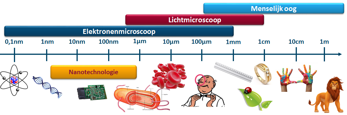
Alle materie rondom ons is opgebouwd uit atomen. Van leeuwen, over haren, tot de chips in je computer. In een gouden ring zitten triljarden atomen. Vele stapjes kleiner komen we de nanodeeltjes tegen, die slechts 100 à 100.000 atomen bevatten. Ze zijn zo klein, dat ze niet langer zichtbaar zijn met het blote oog. Wat maakt deze deeltjes speciaal? En waarom willen we tellen hoeveel atomen ze bevatten? Het antwoord op deze vragen vinden we in hun eigenschappen. Nanodeeltjes bezitten eigenschappen die hun grote varianten niet kunnen evenaren. Maak je goud in het groot, dan krijg je bijvoorbeeld een robuuste goudkleurige ring, die niet reageert met de lucht. Maak je daarentegen minieme goud nanodeeltjes, dan merk je dat deze een heel andere kleur vertonen en bovendien heel sterk chemisch reageren.
De kleine deeltjes bezitten verrassende eigenschappen, erg verschillend van hun grote varianten, en erg gevoelig aan het aantal atomen. Enkele atomen meer of minder tussen de triljarden andere atomen in een gouden ring hebben geen effect op zijn eigenschappen. Enkele atomen meer of minder in een ontzettend klein nanodeeltje daarentegen veranderen zijn kleur, chemische reacties en daarmee ook zijn toepassingen. Door het aantal atomen in de nanodeeltjes te tellen, kennen we de precieze grootte en de innovatieve eigenschappen van het nanodeeltje. Dit is zeer interessant voor gerichte aflevering van medicatie in het menselijk lichaam, kleinere chips in computers en smartphones, lichtere en stevigere materialen, zuivering van de lucht, en talloze andere revolutionaire toepassingen waarop onze moderne maatschappij steunt.
Miljoenen keren ingezoomd
anodeeltjes zijn ontzettend klein, onzichtbaar voor het menselijk oog. Zelfs met een lichtmicroscoop, waarmee je tijdens je middelbare schoolcarrière naar cellen keek tijdens de lessen biologie, kunnen we niet genoeg vergroten om de nanodeeltjes in detail te bekijken! We kunnen ze enkel zien met behulp van een zogenaamde elektronenmicroscoop, een microscoop die miljoenen keren vergroot. Deze vergroting bekomen we door het materiaal te beschijnen met een bundel elektronen, in plaats van met een lichtbundel. De onderzoeksgroep EMAT (Electron Microscopy for Materials Science) van de Universiteit Antwerpen waar ik mijn onderzoek gevoerd heb, beschikt over verschillende elektronenmicroscopen. Het zijn grote, dure machines die met veel zorg gebruikt worden. Dat is ook nodig, want atomen in een nanodeeltje van elkaar onderscheiden met behulp van een elektronenmicroscoop kan je vergelijken met het onderscheiden van twee legoblokjes die op de maan liggen vanuit je achtertuin.
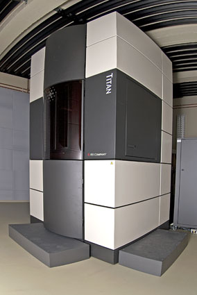 Een microscoop zo groot als een kamer
Verborgen informatie
Een voorbeeld van een foto genomen met een elektronenmicroscoop waarin de atomen zichtbaar zijn, wordt hiernaast getoond. Op de foto zie je enkel de voorkant van het deeltje, net zoals je op een foto van het maanoppervlak ook kraters aan de achterkant van de maan niet ziet liggen.
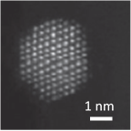 Stevig ingezoomde foto van een nanodeeltje
Een nanodeeltje bestaat uit atoomkolommen, die je kan vergelijken met blokkentorens. Er zitten meerdere blokjes gestapeld in een toren. Het aantal atomen in een nanodeeltje tellen is vergelijkbaar met het tellen van het aantal legoblokjes in een toren die astronauten bouwden op de maan. In een foto zien we niet zomaar hoeveel blokjes er onder elkaar zitten. Deze informatie is verborgen. Om de blokken toch te tellen, wordt daarom een speciale instelling van de microscoop gebruikt, waarmee een hogere toren er lichter uitziet in de foto. Dat helpt ons op weg bij het ontsleutelen van de verborgen informatie.
Oud ...
Om het aantal atomen verborgen in de torens te achterhalen, bestonden reeds twee methoden. De eerste methode vergelijkt de foto’s rechtstreeks met gesimuleerde beelden, wat heel snel telresultaten oplevert. Een belangrijk nadeel aan deze methode is dat we niet weten hoe nauwkeurig deze telling is. De tweede methode gebruikt geavanceerde statistische technieken waarmee het aantal atomen heel nauwkeurig geteld kan worden. Jammer genoeg kunnen we met deze methode het aantal atomen niet tellen op basis van onderbelichte foto’s van ontzettend kleine nanodeeltjes.
… en nieuw
De nieuwe hybride methode die ik ontwikkeld heb combineert de voordelen van de twee reeds bestaande methoden voor het tellen van atomen, om zo hun nadelen te vermijden. Je kan de methode vergelijken met een hybride wagen. Zo’n wagen combineert de voordelen van elektrische wagens met die van de traditionele wagen met verbrandingsmotor. Voor het milieu is een volledig elektrische wagen de betere keuze. Maar je wil toch halverwege je reis niet met een lege batterij stilvallen? De hybride wagen werd daarom ontworpen als een meer betrouwbaar alternatief. De nieuwe hybride methode voor het tellen van atomen is ook een meer betrouwbaar alternatief, waarmee we nu voor het eerst wel atomen kunnen tellen in onderbelichte foto’s.
Moeilijk gaat ook
Je denkt nu waarschijnlijk, als tellen in onderbelichte foto’s dan zo moeilijk is, waarom neem je dan niet gewoon beter belichte foto’s van de nanodeeltjes? Het antwoord is heel eenvoudig. Voor sommige nanodeeltjes gaat dat gewoon niet. Een goed belichte foto bekomen we enkel door veel elektronen op de nanodeeltjes af te vuren, maar dat maakt ze stuk. Het resultaat is dus onvermijdelijk een onderbelichte foto. De deeltjes waar het hier om gaat zijn de allerkleinste nanodeeltjes. Ze helpen bij chemische reacties zoals de afbraak van CO en hebben erg interessante eigenschappen.
 Atomen tellen voor het eerst gelukt in dit ontzettend klein nanodeeltje
Met de bestaande methoden was elke poging tot het tellen van het aantal atomen in deze kleine nanodeeltjes tevergeefs. Met de sterk verbeterde hybride methode is tellen in zo’n moeilijke nanodeeltjes nu wel mogelijk. Moeilijk gaat ook! Dankzij de nieuwe hybride methode zal het onderzoek naar deze interessante, allerkleinste nanodeeltjes nu beter gevoerd kunnen worden. Benieuwd welke nieuwe technologische snufjes ons hierdoor de komende jaren nog te wachten staan!
Bibliografie
- Allen, L., Findlay, S., Oxley, M., and Rossouw, C. (2003). Lattice-resolution contrast from a focused coherent electron probe. Part I. Ultramicroscopy, 96:47–63.
- Anderson, S., Birkeland, C., Anstis, G., and Cockayne, D. (1997). An approach to quantitative compositional profiling at near-atomic resolution using high-angle annular dark field imaging. Ultramicroscopy, 69:83–103.
- Bals, S., Van Aert, S., Romero, C., Lauwaet, K., Van Bael, M., Schoeters, B., Partoens, B., Yücelen, E., Lievens, P., and Van Tendeloo, G. (2012). Atomic scale dynamics of ultrasmall germanium clusters. Nature Communications, 3:897.
- Biernacki, C., Celeux, G., and Govaert, G. (2000). Assessing a mixture model for clustering with the integrated classification likelihood. IEEE Transactions on Pattern Analysis and Machine Intelligence, 22:719–725.
- Bollig, B., Fischer, H., and Kubalek, E. (1996). Multislice simulation of high-resolution scanning transmission electron microscopy Z-contrast images of semiconductor heterointerfaces. Scanning, 18:291–300.
- Boschker, H., Huijben, M., Vailionis, A., Verbeeck, J., Van Aert, S., Luysberg, M., Bals, S., Van Tendeloo, G., Houwman, E., Koster, G., Blank, D., and Rijnders, G. (2011). Optimized fabrication of high-quality La0.67Sr0.33MnO3 thin films considering all essential characteristics. Journal of Physics D: Applied Physics, 44:205001.
- Cowley, J., Hansen, M., and Wang, S. (1995). Imaging modes with an annular detector in STEM. Ultramicroscopy, 58:18–24.
- Cowley, J. and Moodie, A. (1959). The scattering of electrons by atoms and crystals. III. Single-crystal diffraction patterns. Acta Crystallographica, 12:360–367.
- Croitoru, M., Van Dyck, D., Van Aert, S., Bals, S., and Verbeeck, J. (2006). An efficient way of including thermal diffuse scattering in simulation of scanning transmission electron microscopic images. Ultramicroscopy, 106:933–940.
- De Backer, A., Martinez, G., MacArthur, K., Jones, L., Béché, A., Nellist, P., and Van Aert, S. (2015a). Dose limited reliability of quantitative annular dark field scanning transmission electron microscopy for nano-particle atom-counting. Ultramicroscopy, 151:56–61.
- De Backer, A., De wael, A., Gonnissen, J., and Van Aert, S. (2015b). Optimal experimental design for nano-particle atom-counting from high-resolution STEM images. Ultramicroscopy, 151:46–55.
- De Backer, A., Martinez, G., Rosenauer, A., and Van Aert, S. (2013). Atom counting in HAADF STEM using a statistical model-based approach: Methodology, possibilities, and inherent limitations. Ultramicroscopy, 134:23–33.
- Dempster, A., Laird, N., and Rubin, D. (1977). Maximum Likelihood from Incomplete Data via the EM Algorithm. Journal of the Royal Statistical Society. Series B (Methodological), 39:1–38.
- den Dekker, A., Van Aert, S., van den Bos, A., and Van Dyck, D. (2005). Maximum likelihood estimation of structure parameters from high resolution electron microscopy images. Part I: a theoretical framework. Ultramicroscopy, 104:83106.
- E, H., MacArthur, K., Pennycook, T., Okunishi, E., D’Alfonso, A., Lugg, N., Allen, L., and Nellist, P. (2013). Probe integrated scattering cross sections in the analysis of atomic resolution HAADF STEM images. Ultramicroscopy, 133:109119.
- Erni, R., Rossell, M., Kisielowski, C., and Dahmen, U. (2009). Atomic-Resolution Imaging with a Sub-50-pm Electron Probe. Physical Review Letters, 102:096101.
- Fertig, J. and Rose, H. (1981). Resolution and contrast of crystalline objects in high-resolution scanning-transmission electron-microscopy. Optik, 59:407–429.
- Findlay, S., Allen, L., Oxley, M., and Rossouw, C. (2003). Lattice-resolution contrast from a focused coherent electron probe. Part II. Ultramicroscopy, 96:65–81.
- Findlay, S. and LeBeau, J. (2013). Detector non-uniformity in scanning transmission electron microscopy. Ultramicroscopy, 124:52–60.
- Fujita, H. and Sumida, N. (1994). Physics of New Materials - Chapter 8 Usefulness of Electron Microscopy. New York: Springer Science+Business Media LLC.
- Geuens, P. and Van Dyck, D. (2005). The S-State Model for Electron Channeling in High-Resolution Electron Microscopy. Advances in Imaging and Electron Physics, 136:111–226.
- Grieb, T., Müller, K., Fritz, R., Schowalter, M., Neugebohrn, N., Knaub, N., Volz, K., and Rosenauer, A. (2012). Determination of the chemical composition of GaNAs using STEM HAADF imaging and STEM strain state analysis. Ultramicroscopy, 117:15–23.
- Grillo, V. (2011). An advanced study of the response of ADF detector. Journal of Physics: Conference Series, 326:012036.
- Haider, M., Uhlemann, S., Schwan, E., Rose, H., Kabius, B., and Urban, K. (1998). Electron microscopy image enhanced. Nature, 392:768–769.
- Hawkes, P., Spence, J., and Nellist, P. (2007). Science of Microscopy - Chapter 2 Scanning Transmission Electron Microscopy. New York: Springer Science+Business Media LLC.
- Henderson, R. (1995). The potential and limitations of neutrons, electrons and X-rays for atomic resolution microscopy of unstained biological molecules. Q Rev Biophys, 28:171–193.
- Huang, T. and Nancy Xu, X. (2010). Synthesis and Characterization of Tunable Rainbow Colored Colloidal Silver Nanoparticles Using Single-Nanoparticle Plasmonic Microscopy and Spectroscopy. Journal of Materials Chemistry, 20:9867–9876.
- Huijben, M., Koster, G., Kruize, M., Wenderich, S., Verbeeck, J., Bals, S., Slooten, E., Shi, B., Molegraaf, H., Kleibeuker, J., Van Aert, S., Goedkoop, J., Brinkman, A., Blank, D., Golden, M., Van Tendeloo, G., Hilgenkamp, H., and Rijnders, G. (2013). Defect Engineering in Oxide Heterostructures by Enhanced Oxygen Surface Exchange. Advanced Functional Materials, 24:5240.
- Ishizuka, K. (2002). A practical approach for STEM image simulation based on the FFT multislice method. Ultramicroscopy, 90:71–83.
- Jones, L. (2016). Quantitative ADF STEM: acquisition, analysis and interpretation. IOP Conf. Series: Materials Science and Engineering, 109:012008.
- Jones, L., MacArthur, K., Fauske, V., van Helvoort, A., and Nellist, P. (2014). Rapid estimation of catalyst nanoparticle morphology and atomic-coordination by high-resolution Z-contrast electron microscopy. Nano Letters, 14:6336–6341.
- Jones, L. and Nellist, P. (2013). Identifying and Correcting Scan Noise and Drift in the Scanning Transmission Electron Microscope. Microscopy and Microanalysis, 19:1050–1060.
- Jones, L., Yang, H., Pennycook, T., Marshall, M., Van Aert, S., Browning, N., Castell, M., and Nellist, P. (2015). Smart Align a new tool for robust non-rigid registration of scanning microscope data. Advanced Structural and Chemical Imaging, 1:8.
- Kirkland, E. (1998). Advanced computing in electron microscopy. New York: Springer Science+Business Media LLC.
- Koch, C. (2002). Determination of core structure periodicity and point defect density along dislocations. Tempe, AZ: Arizona State University.
- Koli, D., Agnihotri, G., and Purohit, R. (2014). A Review on Properties, Behaviour and Processing Methods for Al- Nano Al2O3 Composites. Procedia Materials Science, 3rd International Conference on Materials Processing and Characterisation (ICMPC 2014), 6:567589.
- LeBeau, J., Findlay, S., Allen, L., and Stemmer, S. (2008). Quantitative Atomic Resolution Scanning Transmission Electron Microscopy. Physical Review Letters, 100:206101.
- LeBeau, J., Findlay, S., Allen, L., and Stemmer, S. (2010). Standardless atom counting in scanning transmission electron microscopy. Nanoletters, 10:4405–4408.
- Lin, W., Insley, T., Tuttle, M., Zhu, L., Berthold, D., Kràl, P., Rienstra, C., and Murphy, C. (2015). Control of Protein Orientation on Gold Nanoparticles. The Journal of Physical Chemistry C, 119:2103521043.
- Loane, R., Xu, P., and Silcox, J. (1991). Thermal vibrations in convergent-beam electron diffraction. Acta Crystallographica Section A, 47:267–278.
- MacArthur, K., D'Alfonso, A., Ozkaya, D., Allen, L., and Nellist, P. (2015). Optimal ADF STEM imaging parameters for tilt-robust image quantification. Ultramicroscopy, 156:1–8.
- MacArthur, K., Jones, L., and Nellist, P. (2014). How flat is your detector? Non-uniform annular detector sensitivity in STEM quantification. Journal of Physics: Conference Series, 522:012018.
- Martinez, G. (2014). Quantitative Model-based High Angle Annular Dark Field Scanning Transmission Electron Microscopy. PhD thesis, University of Antwerp.
- Martinez, G., De Backer, A., Rosenauer, A., Verbeeck, J., and Van Aert, S. (2013). The effect of probe inaccuracies on the quantitative model-based analysis of high angle annular dark field scanning transmission electron microscopy images. Micron, 63:57–63.
- Martinez, G., Jones, L., De Backer, A., Béché, A., Verbeeck, J., Van Aert, S., and Nellist, P. (2015). Quantitative STEM normalisation: The importance of the electron flux. Ultramicroscopy, 159:46– 58.
- McLachlan, G. and Peel, D. (2000). Finite Mixture Models. Wiley series in probability and statistics. John Wiley and Sons, inc.
- Mehrtens, T., Schowalter, M., Tytko, D., Choi, P., Raabe, D., Hoffmann, L., J¨onen, H., Rossow, U., Hangleiter, A., and Rosenauer, A. (2013). Measurement of the indium concentration in high indium content InGaN layers by scanning transmission electron microscopy and atom probe tomography. Applied Physics Letters, 102:132112.
- Meyer, J., Kotakoski, J., and Mangler, C. (2014). Atomic structure from large-area, low-dose exposures of materials: A new route to circumvent radiation damage. Ultramicroscopy, 145:13–21.
- Mikami, Y., Dhakshinamoorthy, A., Alvaro, M., and Garc´ıa, H. (2013). Catalytic activity of unsupported gold nanoparticles. Catalysis Science & Technology, 3:58–69.
- Moore, G. (1998). Cramming More Components onto Integrated Circuits. Proceedings of the IEEE, 86:82–85.
- Muller, D., Edwards, B., Kirkland, E., and Silcox, J. (2001). Simulation of thermal diffuse scattering including a detailed phonon dispersion curve. Ultramicroscopy, 86:371–380.
- Nellist, P. and Pennycook, S. (1999). Incoherent imaging using dynamically scattered coherent electrons. Ultramicroscopy, 78:111–124.
- Nellist, P. and Pennycook, S. (2000). The Principles and Interpretation of Annular Dark-Field ZContrast Imaging. Advances in imaging and electron physics, 113:147.
- Nguyen, D., Findlay, S., and Etheridge, J. (2014). The spatial coherence function in scanning transmission electron microscopy and spectroscopy. Ultramicroscopy, 146:6–16.
- Olson, G. (2000). Pathways of Discovery, Designing a New Material World. Science, 288:993–998.
- Ralph, T. and Hogarth, M. (2002). Catalysis for Low Temperature Fuel Cells. Platinum Metals Review, 46:117–135.
- Rosenauer, A., Mehrtens, T., Mu¨ller, K., Gries, K., Schowalter, M., Satyam, P., Bley, S., Tessarek, C., Hommel, D., Sebald, K., Seyfried, M., Gutowski, J., Avramescu, A., Engl, K., and Lutgen, S. (2011). Composition mapping in InGaN by scanning transmission electron microscopy. Ultramicroscopy, 111:1316–1327.
- Rosenauer, A. and Schowalter, M. (2007). STEMSIM - A new software tool for simulation of STEM HAADF Z-contrast imaging. Springer Proceedings in Physics, 120:169.
- Rosenauer, A., Schowalter, M., Titantah, J., and Lamoen, D. (2008). An emission-potential multislice approximation to simulate thermal diffuse scattering in high-resolution transmission electron microscopy. Ultramicroscopy, 108:1504–1513.
- Ruskin, R., Yu, Z., and Grigorieff, N. (2013). Quantitative characterization of electron detectors for transmission electron microscopy. Journal of Structural Biology, 184:385393.
- Shah, A., Rahman, L., Qureshi, R., and Rehman, Z. (2012). Synthesis, characterization and applications of bimetallic (Au-Ag, Au-Pt, Au-Ru) alloy nanoparticles. Reviews on Advanced Materials Science, 30:133–149.
- Shibata, N., Kohno, Y., Findlay, S., Sawada, H., Kondo, Y., and Ikuhara, Y. (2010). New area detector for atomic-resolution scanning transmission electron microscopy. Journal of Electron Microscopy, 59:473–479.
- Spence, J. (1999). The future of atomic resolution electron microscopy for materials science. Materials Science and Engineering: R: Reports, 26:1–49.
- Stone, J., Jackson, S., and Wright, D. (2011). Biological applications of gold nanorods. Wiley Interdiscip Rev Nanomed Nanobiotechnol., 3:100–109.
- Takayanagi, K., Kim, S., Lee, S., Oshima, Y., Tanaka, T., Tanishiro, Y., Sawada, H., Hosokawa, F., Tomita, T., Kaneyama, T., and Kondo, Y. (2011). Electron microscopy at a sub-50 pm resolution. Journal of Electron Microscopy, 60:239–244.
- Toshima, N. and Yonezawa, T. (1998). Bimetallic nanoparticlesnovel materials for chemical and physical applications. New Journal of Chemistry, 22:1179–1201.
- Van Aert, S., Batenburg, K., Rossell, M., Erni, R., and Van Tendeloo, G. (2011). Three-dimensional atomic imaging of crystalline nanoparticles. Nature, 470:374–377.
- Van Aert, S., De Backer, A., Martinez, G., Goris, B., Bals, S., and Van Tendeloo, G. (2013). Procedure to count atoms with trustworthy single-atom sensitivity. Physical Review B, 87:064107.
- Van Aert, S., den Dekker, A., van den Bos, A., and Van Dyck, D. (2005). Maximum likelihood estimation of structure parameters from high resolution electron microscopy images. Part II: a practical example. Ultramicroscopy, 104:107125.
- Van Aert, S., Turner, S., Delville, R., Schryvers, D., Van Tendeloo, G., and Salje, E. (2012). Direct Observation of Ferrielectricity at Ferroelastic Domain Boundaries in CaTiO3 by Electron Microscopy. Advanced materials, 24:523.
- Van Aert, S., Verbeeck, J., Erni, R., Bals, S., Luysberg, M., Van Dyck, D., and Van Tendeloo, G. (2009). Quantitative atomic resolution mapping using high-angle annular dark field scanning transmission electron microscopy. Ultramicroscopy, 109:12361244.
- van den Bos, K., De Backer, A., Martinez, G., Winckelmans, N., Bals, S., Nellist, P., and Van Aert, S. (2016). Unscrambling mixed elements using high angle annular dark field scanning transmission electron microscopy. Physical Review Letters, accepted.
- van den Bos, K. and Van Aert, S. (2014). A channelling based approach for scattering cross sections of mixed columns in HAADF STEM images. 18th International Microscopy Congress IMC 2014, Prague, Czech Republic, pages IT–2–P–2255.
- Van Dyck, D., Van Aert, S., den Dekker, A., and van den Bos, A. (2003). Is atomic resolution transmission electron microscopy able to resolve and refine amorphous structures? Ultramicroscopy, 98:27–42.
- Vigderman, L., Khanal, B., and Zubarev, E. (2012). Functional Gold Nanorods: Synthesis, SelfAssembly, and Sensing Applications. Advanced Materials, 24:48114841.
- Wiesendanger, R. (1994). Scanning Probe Microscopy and Spectroscopy, Methods and Applications. Cambridge: Cambridge University Press.
- Yang, H., Pennycook, T., and Nellist, P. (2015). Efficient phase contrast imaging in STEM using a pixelated detector. Part II: Optimisation of imaging conditions. Ultramicroscopy, 151:232239.
- Yang, P. and Tarascon, J. (2012). Towards systems materials engineering. Nature Materials, 11:560– 563.
- Zaleska-Medynska, A., Marchelek, M., Diak, M., and Grabowska, E. (2016). Noble metal-based bimetallic nanoparticles: the effect of the structure on the optical, catalytic and photocatalytic properties. Advances in Colloid and Interface Science, 229:80–107.
- Zanchet, D., Hall, B., and Ugarte, D. (2000). Characterization of Nanophase Materials - Chapter 2 X-ray Characterization of Nanoparticles. Weinheim: Wiley-VCH.